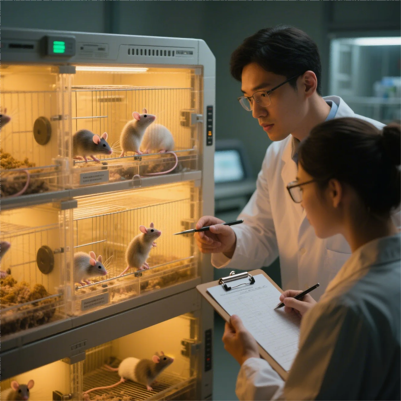
Variability enters a cell-derived xenograft (CDX) study on a timeline. It may begin during cell expansion, widen at implantation, shift again at enrollment, and become visible only when tumor curves separate. A well-run cdx model experiment tracks that timeline so biological heterogeneity remains visible while avoidable differences in materials, handling, dosing, and measurement are contained.
The most useful planning tool is a variability map. Biological inputs form one category, establishment and treatment operations form another, and measurement plus analysis form a third. Each category can be linked to controls, acceptance limits, and records before animals are enrolled.
Real tumor heterogeneity belongs in the data. Overcontrol can create a system that no longer answers the scientific question, while loose operations bury the biological response in preventable noise. The variability map separates those two forms of spread and names the person responsible for watching each controllable input.
Historical data help set realistic expectations. Tumor-take rate, growth distribution, time to enrollment, control-group variance, and prior drug sensitivity can inform cohort size and scheduling. Those records are most useful when cell bank, passage range, host strain, implantation method, and measurement rules remain traceable. The team revisits control limits when a new bank, host source, or procedure materially changes performance.
Control Biological Inputs Before Implantation
Methods such as short tandem repeat (STR) profiling confirm cell identity before implantation, with mycoplasma and relevant pathogen testing used to protect model integrity. Authentication is not a one-time administrative step; testing points reflect banking, expansion, transport, and the risk of contamination or misidentification.
The team quantifies residual variation so the analytical plan can accommodate it honestly. A well-characterized CDX model begins with a cell source whose identity and condition are demonstrable. Together, these checks make cell preparation a controlled input rather than an assumption.
Culture status influences tumor establishment. Passage number, confluence, viability, media, serum lot, harvest method, and time between preparation and injection remain within predefined ranges. A standardized worksheet can capture these details and prevent a rushed preparation from becoming an unexplained difference in tumor growth.
Model-resource depth can support more consistent CDX planning when the underlying cell and tumor histories are well documented. Jennio Biotech‘s platform includes more than 1,000 validated tumor cell lines and 500+ CDX models, giving sponsors a broader base for matching cancer type, molecular background, and study objective. Project-specific controls should still define cell preparation, implantation, monitoring, and deviation handling before the study begins.
Model selection belongs in the same control strategy. Cancer type, target expression, mutation profile, baseline growth, and known sensitivity or resistance determine relevance. A slower model with a clear mechanism may generate a more useful answer than a fast-growing line chosen solely for operational convenience.
Standardize Establishment, Grouping, and Dosing
Implantation uses a defined site, needle path, depth, volume, and trained operator technique. Leakage, misplaced inoculum, or tissue trauma can change take and growth. Competency records and periodic observation of the procedure help distinguish operator drift from biological change. Cryopreserved master and working banks can limit genetic and phenotypic drift across campaigns. For CDX studies, the enrollment window can be treated as a controlled experimental variable when tumor growth criteria are part of the study design.
The team fixes enrollment criteria before tumors appear. Minimum and maximum volume, acceptable growth pattern, health status, and enrollment window can prevent selective inclusion. Groups are then balanced by baseline tumor volume and relevant animal characteristics, using a documented randomization method rather than visual judgment.
Dosing introduces another set of variables. The protocol fixes formulation preparation, concentration verification, route, volume, timing, sequence, and operator assignment. If different groups require different handling, the order can rotate so that time of day and preparation age do not become hidden treatment effects. The team rejects a cell preparation when viability or handling time falls outside the agreed acceptance range and rejects a dosing preparation when concentration, stability, or preparation time falls outside its predefined limits.
Throughout the study, staff record body weight, tumor measurements, clinical condition, dose administration, and deviations on schedule. The same early-removal criteria apply to every group. The data history retains each missed dose or ulcerated tumor, and a prespecified analytical rule governs its treatment.
Reduce Noise During Measurement and Analysis
Caliper measurements require a consistent orientation, technique, and tumor-volume formula. The same trained assessor or blinded assessors can reduce reader variation. Periodic duplicate measurements reveal drift, while imaging can support deeply seated, irregular, or otherwise difficult lesions when the study design justifies it. Pilot qualification can confirm take rate and growth behavior after a material or site change.
No single endpoint carries the entire interpretation. Terminal tumor weight, longitudinal volume, imaging, pathology, immunohistochemistry, and molecular biomarkers can test different aspects of response. Agreement among them helps distinguish genuine antitumor activity from edema, necrosis, measurement geometry, or a transient growth delay.
Before database lock, rules define outliers, missing observations, early termination, non-takes, and protocol deviations. Statistical methods need to match repeated measurements and group structure. Keeping raw caliper records, images, pathology slides, analysis settings, and decision logs allows an independent reviewer to reconstruct the result. Recorded proficiency is especially important when several technicians share implantation work.
Variability never disappears from a living tumor study, nor should it. The objective is to know where the remaining spread comes from. Once cell history, implantation, enrollment, dosing, measurement, and analysis are documented on the same timeline, the next CDX campaign can reproduce the method—or explain precisely why it changed.